


Fibroids
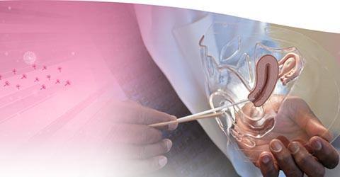
Uterine fibroids are non-cancerous (benign) tumours, commonly seen in women of childbearing age. Fibroids are composed of muscle cells and other tissues. They develop in and around the wall of the uterus or womb. Uterine fibroids are usually round or semi-round in shape. Based on their location within the uterus, uterine fibroids can be classified as:
- Subserosal fibroids: Sited beneath the serosa (the membrane covering the outer surface of the uterus)
- Submucosal fibroids: Sited inside the uterine cavity below the inside layer of the uterus
- Intramural fibroids: Sited within the muscular wall of the uterus
- Intracavitary fibroids: Sited inside the uterine cavity
- Pedunculated fibroids: Develop on a stalk attached to the outer wall of the uterus
Causes
The exact cause for the development of fibroids remains unknown, but some of the proposed causes include:
- Genetic abnormalities
- Alterations in expression of growth factor (protein involved in rate and extent of cell proliferation)
- Abnormalities in the vascular system
- Tissue response to injury
- Family history of fibroids
- Uterine infection
- Consumption of alcohol
- Elevated blood pressure
- Hormonal imbalance during puberty
Symptoms
The majority of women with uterine fibroids may be asymptomatic. However the basic symptoms associated with fibroids include:
- Heavy menstrual bleeding
- Prolonged menstrual periods
- Pelvic pressure or pain
- Frequent urination
- Constipation
- Backache or leg pain
- Difficulty in emptying your bladder
Diagnosis
The diagnosis of uterine fibroids involves a pelvic examination, followed by ultrasound evaluation. Other imaging techniques such as MRI scan and CT scan may also be employed.
Treatment
Different methods are being used for managing uterine fibroids. Surgery is considered the best modality of treatment. The common surgeries performed for the management of fibroids include:
- Hysterectomy or removal of the uterus
- Myomectomy or selective removal of the fibroids within the uterus
- Destructive techniques that involve boring holes into the fibroids with a laser or freezing probes (cryosurgery)
- Other techniques employed are uterine artery embolization (UAE) and uterine artery occlusion (UAO)
Non-surgical methods comprising of steroidal medication are also used to stabilize the oestrogen levels in the body.
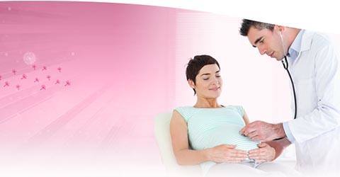
Risks during pregnancy
Some studies indicate that the presence of uterine fibroids during pregnancy increases the risk of complications such as first trimester bleeding, breech presentation, placental abruption, increased chance of caesarean section and problems during labour.